What is PRFM and LPRF? Understanding the Differences in Autologous Blood Therapies
Introduction to PRF and Its Variations
Platelet-rich fibrin (PRF) is an advanced form of autologous blood therapy that enhances tissue regeneration and healing. Two key variations of PRF are:
L-PRF (Leukocyte- and Platelet-Rich Fibrin)
PRFM (Platelet-Rich Fibrin Matrix)
Both are derived from the patient’s own blood but differ in preparation methods, structure, and clinical applications. This article explores what PRFM and LPRF are, their treatment processes, required materials (such as PRF tubes, centrifuges, and calcium chloride tubes), and their key differences.
What is LPRF? (Leukocyte- and Platelet-Rich Fibrin)
Definition & Composition
LPRF is a second-generation platelet concentrate that contains platelets, leukocytes (white blood cells), and fibrin in a dense, three-dimensional matrix. Unlike PRP (platelet-rich plasma), LPRF does not require anticoagulants or additives, making it a fully natural biomaterial.
LPRF Treatment Process
Blood Collection:
Blood is drawn into PRF tubes (no anticoagulant).
Centrifugation:
Spun at low speed (around 2700 RPM for 12-14 minutes) to form a fibrin clot.
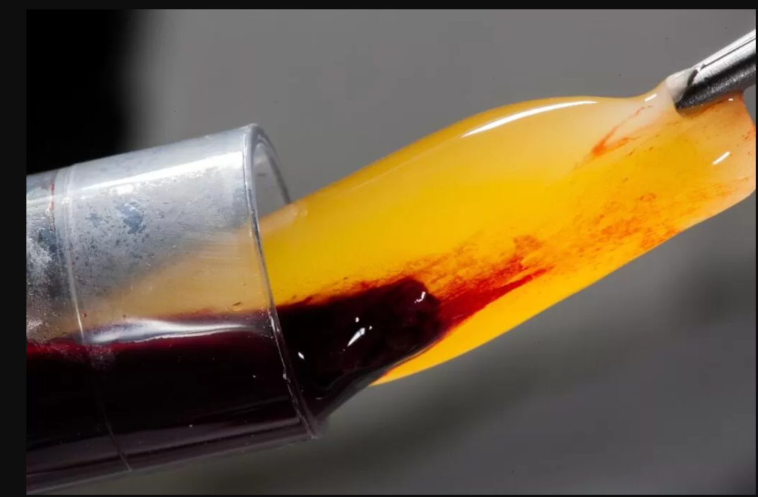
Clot Extraction:
The fibrin clot is separated from red blood cells and compressed into a membrane or plug.
Materials Needed for LPRF
PRF tubes (glass or plastic, no anticoagulant)
Centrifuge (specific PRF protocols)
Surgical tools (to handle fibrin clot)
Clinical Applications of LPRF
✔ Dental surgery (bone grafts, socket preservation)
✔ Wound healing (chronic ulcers, post-surgical recovery)
✔ Orthopedics (tendon repair, osteoarthritis)
What is PRFM? (Platelet-Rich Fibrin Matrix)
Definition & Composition
PRFM is a modified version of PRF that includes additional activation agents (such as calcium chloride or thrombin) to accelerate fibrin polymerization. It forms a stronger, more elastic matrix than traditional PRF.
PRFM Treatment Process
Blood Collection:
Blood is drawn into PRF tubes (sometimes with calcium chloride activation).
Centrifugation:
Spun at controlled speeds (varies by protocol).
Fibrin Activation:
Mixed with calcium chloride tubes to enhance fibrin formation.
Matrix Formation:
The fibrin matrix is extracted and shaped for application.
Materials Needed for PRFM
PRF tubes (sometimes with anticoagulant for delayed activation)
Calcium chloride tube (for fibrin acceleration)
Centrifuge (optimized for PRFM protocols)
Clinical Applications of PRFM
✔ Facial rejuvenation (dermal fillers, collagen stimulation)
✔ Hair restoration (injections for hair growth)
✔ Soft tissue repair (ligament and cartilage regeneration)
Key Differences Between LPRF and PRFM
| Factor | LPRF | PRFM |
| Preparation | No anticoagulants/additives | May use calcium chloride/thrombin |
| Fibrin Structure | Dense, natural fibrin | More elastic, reinforced matrix |
| Centrifugation Speed | Low-speed (2700 RPM) | Variable (depends on protocol) |
| Activation Time | Slow, natural clotting | Faster due to additives |
| Primary Uses | Dental, orthopedic surgery | Aesthetics, hair restoration |
1. Preparation & Additives
LPRF is purely autologous (no additives).
PRFM may use calcium chloride tubes for faster fibrin formation.
2. Fibrin Matrix Strength
LPRF has a denser, more rigid structure (ideal for surgical applications).
PRFM is more flexible (better for injectable treatments).
3. Clinical Applications
LPRF is preferred in dental and orthopedic surgeries where structural integrity is key.
PRFM is used in cosmetic and regenerative medicine where malleability is needed.
Conclusion: Choosing Between LPRF and PRFM
Both LPRF and PRFM are highly effective autologous blood therapies, but their best use depends on the medical or aesthetic application:
For surgical and dental procedures → LPRF (stronger fibrin structure).
For injectable treatments (skin, hair, soft tissue) → PRFM (more flexible matrix).
Understanding the differences in PRF tubes, centrifugation protocols, and activation methods helps clinicians select the best option for optimal patient outcomes.